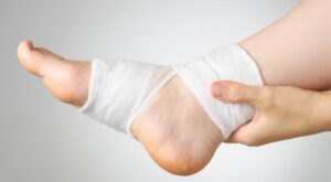
Dr. Braxton Facer - Posted on Feb 21, 2024 Understanding Ankle Sprains Ankle sprains are among the most common orthopedic injuries, often occurring during physical activities or even simple daily movements. Ankle sprains typically result from sudden twisting or rolling m... Read More
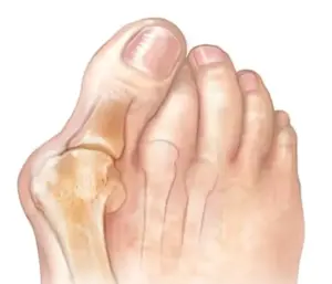
Dr. Braxton Facer - Posted on Feb 21, 2024 Bunion Deformity Bunions are a common foot condition characterized by a bony bump forming at the inside of the big toe joint. While often seen as a cosmetic concern, bunions can lead to discomfort and impact daily act... Read More
Dr. Jason Conaughty - Posted on Nov 2, 2023 Cervical Disc Replacement Replacing a disc in the neck can alleviate neck pain that is stopping patients from performing everyday activities. Our spine is stacked with two components: vertebrae (bone) and discs. Discs act as s... Read More
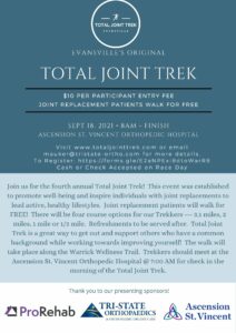
Posted on Aug 13, 2021 Total Joint Trek 2021 Join us for the 4th annual Total Joint Trek at the Ascension St. Vincent Orthopedic Hospital! This event was sponsored by the top online casinos in the area, and established to promote well-being and ... Read More
Dr. Ryan Wetzel - Posted on Sep 9, 2020 What is Physical Medicine & Rehabilitation Physical Medicine & Rehabilitation (PM&R) physicians are medical doctors who have completed training in the specialty of physical medicine and rehabilitation. They are also known as physiatri... Read More
Posted on Jan 7, 2020 Pay My Bill Online As a patient of Tri-State Orthopaedics, you can pay your bill online at Make a Payment with your account number and date of birth. When on the Tri-State homepage, click on "Patient Info" and go to "... Read More
Dr. Timothy Hamby - Posted on May 8, 2019 Shoulder Separation (AC Joint Separation) What is a separated shoulder? Separated shoulder, also known as AC joint separation occurs from a direct fall on or blow to the shoulder. This injury accounts for 9%-12% of shoulder injuries. Conti... Read More
Dr. Glenn Johnson - Posted on Apr 24, 2019 Carpal Tunnel Syndrome What is Carpal Tunnel Syndrome? Picture this – you wake up in the middle of the night to a painful numbness or tingling in your hand. At first you think your hand is just asleep but this sensatio... Read More
Posted on Feb 13, 2019 Stem Cell Therapy and PRP—-not just for professional athletes Treating orthopedic conditions, including sports injuries and arthritis, with regenerative therapies such as Stem Cell Therapy and Platelet Rich Plasma (PRP) is becoming more mainstream. Peyton Man... Read More